
tgoop.com/AdisonInstitute/4407
Last Update:
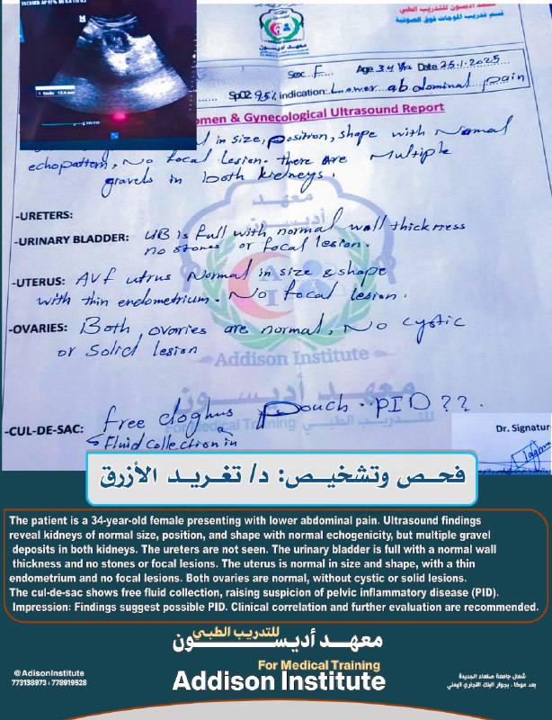
🔰فحص وتشخيص 👩⚕ د/ تغريد الازرق 🔰
➖➖➖➖➖➖➖➖
The patient is a 34-year-old female presenting with lower abdominal pain
♦️Ultrasound Findings:
Ultrasound findings reveal kidneys of normal size, position, and shape with normal echogenicity, but multiple gravel deposits in both kidneys
The ureters are not seen
The urinary bladder is full with a normal wall thickness and no stones or focal lesions
The uterus is normal in size and shape, with a thin endometrium and no focal lesions
Both ovaries are normal, without cystic or solid lesions
The cul-de-sac shows free fluid collection, raising suspicion of pelvic inflammatory disease (PID)
Findings suggest possible PID
Clinical correlation and further evaluation are recommended
🔰للحجز والتسجيل في الدفعات القادمة من السونار 👈 773138973 او 778919528
؛➖➖➖➖
✅لمتابعتنا على👇
♦️قناة Telegram خاصة بالسونار👇
https://www.tgoop.com/UltrasoundAddison
♦️قناة اديسونTelegram👇
https://www.tgoop.com/AdisonInstitute
♦️مجتمع واتساب خاص بالسونار 👇
https://chat.whatsapp.com/IGvnrUm0GBmFZjdVhYtv4F
BY معهد اديسون Addison institute
Share with your friend now:
tgoop.com/AdisonInstitute/4407